Category: Uncategorized
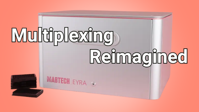
Meet EYRA: Multiplexing Reimagined
A Joe Blogs post by Joe Roberts, PhD
When it comes to multiplex protein analysis, researchers need accuracy, speed, and simplicity. Traditional flow-based systems have long been the standard, but they bring challenges: sheath fluids, blocked probes, and constant maintenance. Enter the Mabtech EYRA™ – a fluidics-free multiplex immunoassay platform that reimagines how scientists generate cytokine and biomarker data. With confocal imaging, RAWsphere analysis, and compatibility with EYRAplex bead kits, EYRA makes multiplexing faster, simpler, and more reliable.
Multiplex Without Compromise
With EYRAplex magnetic bead assays, EYRA can quantify more than 30 analytes from a single sample – whether that’s serum, plasma, or cell culture supernatant. This means less sample consumption, fewer runs, and richer datasets for every experiment.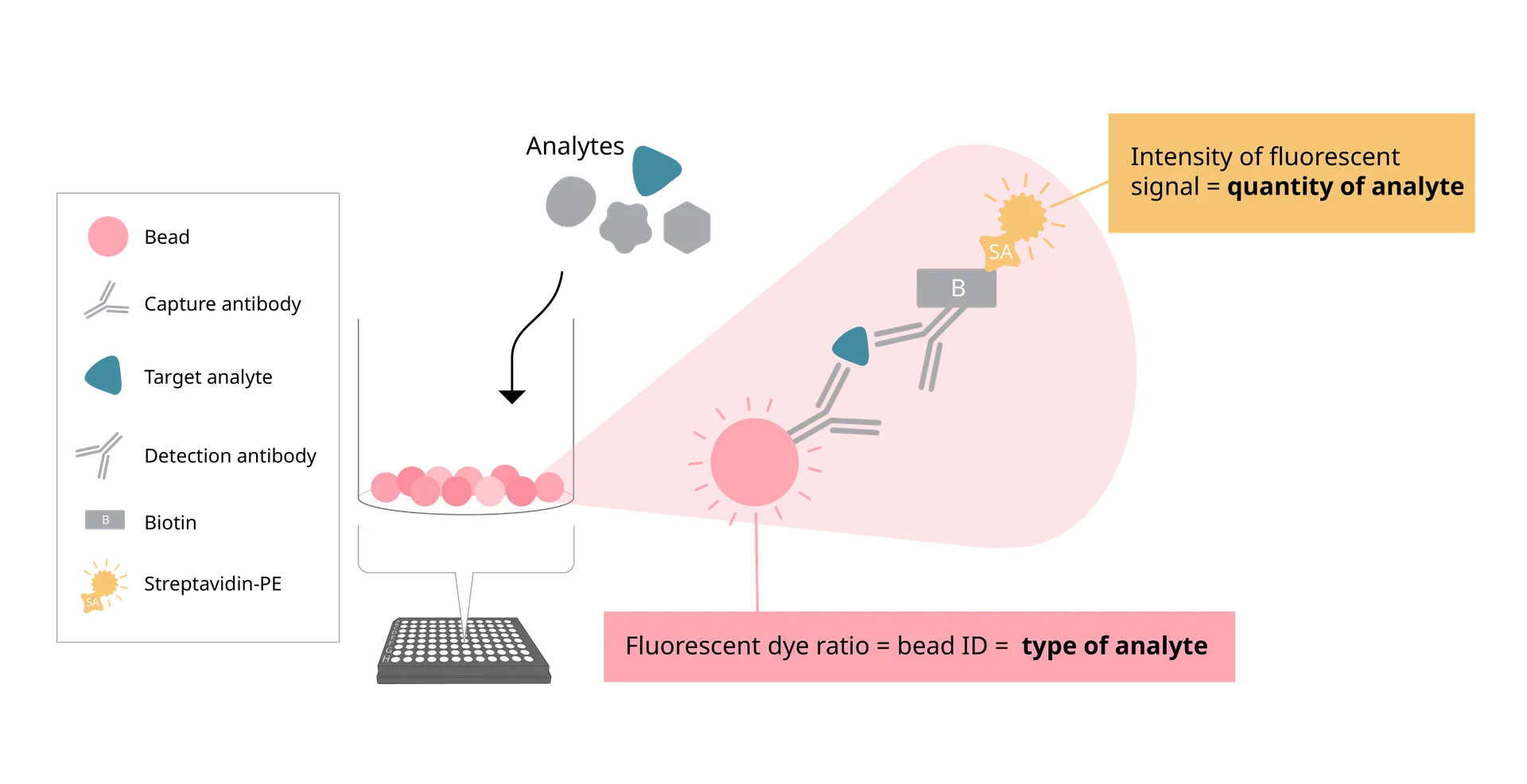
At the core of EYRA’s precision is the RAWsphere image analysis algorithm. It identifies each bead, links it to the correct analyte, and quantifies the PE signal with high resolution. Whether you’re measuring six cytokines or a full 30+ panel, RAWsphere ensures your multiplex data is accurate and reproducible.
Watch video to see how EYRA works
Fluidics-Free Technology
EYRA is built around a completely flow-free design. No sheath fluid, priming, waste handling or blocked probes.
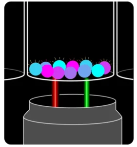
Instead of pushing samples through fluidics, EYRA uses confocal microscopy to image settled magnetic beads directly in the wells of a 96-well plate. Each bead carries a unique fluorescent dye signature for identification, while analyte-bound PE-labelled antibodies provide the quantitative signal.
The result? You insert your plate, select your assay in the intuitive Mabtech Opal™ software, and hit read. Fifteen minutes later, you have fully processed data – with your samples never leaving the wells.
From Plate to Excel - Fast
Opal™ software comes preloaded with templates for every EYRAplex kit. Standards, plate layouts, and gating are handled automatically – so you spend less time setting up and more time on results.
Once the plate read is complete, results are exported directly into Excel, with options for bulk export if you’re working on large studies or multiple plates. No manual reformatting. No data wrangling headaches.
The Maintenance-Free Mindset
Because EYRA is fluidics-free, there’s no daily calibration or cleaning. No flushing, no wasted consumables, and no time lost to instrument downtime. It’s genuinely a plug-and-play experience – switch it on, run your plate, and walk away with your data.
Joe’s Takeaway
The Mabtech EYRA isn’t just another multiplex platform – it’s multiplexing reimagined. By removing fluidics and harnessing high-resolution confocal imaging, EYRA delivers:
- High-plex capacity: 30+ analytes per well
- Fast turnaround: ~15 minutes per plate
- No maintenance: no daily cleaning or calibration
- Accurate, reproducible results: powered by RAWsphere analysis
For labs looking to streamline their workflow without sacrificing data quality, EYRA offers a fresh, frustration-free alternative to traditional flow-based systems.
Until next time… happy experimenting!
Joe Roberts, PhD
Product Manager
Millennium Science

10x Genomics Grant Application Resources
Why 10x Genomics?
10x Genomics is a proven leader in single cell research, expanding access to single cell with optimised protocols, end-to-end support, and easy-to-use analysis tools. 10x Genomics’ technology – backed by 10 years, over 2,200 patents, and > $1.5B in R&D investment – has empowered researchers to publish over 8,000 studies, including many high-impact applications of their tools.
10x Genomics’ complementary Visium and Xenium platforms offer an essential balance between unbiased discovery and precision insights and are leading the way in performance, flexibility, and ease-of-use. Being at the nexus of spatial biology means seeing what others may have missed: global cell-type organisation, cell–cell interactions, heterogeneous spatial niches, novel gene programs, critical ligand–receptor signalling networks, and spatial biomarkers of therapeutic response.
Download grant application resources here:
Enhance your chances of obtaining research funding for your Single Cell and Spatial Transcriptomics projects with the below 10x Genomics grant application resources.


These grant application resources include:
- Technology summaries
- Applications
- Workflow overviews
- Technology Advantages
- Validation studies & data benchmarking
- Data processing & analysis
- Sample & project size considerations
- Additional resources
- References
Other useful resources:
Book a project discussion or request a quote below:
Deeper Spatial Discovery with Visium HD 3′
Visium HD 3' is here! Explore more species - Now Shipping
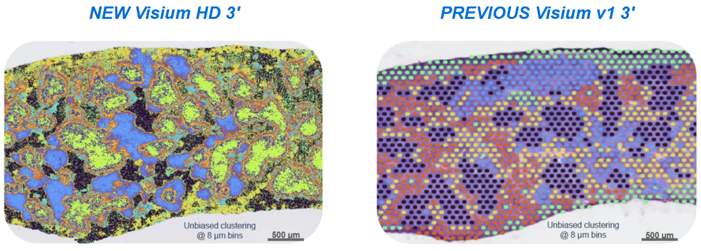
Introducing 10x Genomics‘ Visium HD 3’, a novel assay for unbiased cross-species spatial gene expression profiling of fresh frozen tissue sections mounted on a standard glass slide. The Visium HD’s array has a gapless design that enables integration of unsupervised gene expression clustering data with microscope H&E images from the same tissue section, allowing precise profiling of finer anatomical features with high tissue coverage at single cell-scale resolution.
Furthermore, this novel reverse transcription-based assay generates cDNA products compatible with both short reads and long reads sequencing, extending its applications beyond gene expression analysis to enable immune profiling and the discovery of isoforms and novel transcripts on a spatial level.

Key Features
Visium HD 3’ assay, which delivers whole transcriptome spatial analysis with a 3’ poly(A) capture-based chemistry. This assay provides:
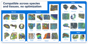
- Flexibility to explore the spatial biology of fresh frozen samples from any species
- De novo spatial discovery power, including isoforms, TCRs/BCRs, and SNVs
- True single cell resolution with new cell segmentation software
- Streamlined slide handling and high spatial fidelity powered by the Visium CytAssist instrument
Space Ranger v4.0.1 is now live! Expand the power of Visium HD with cell segmentation
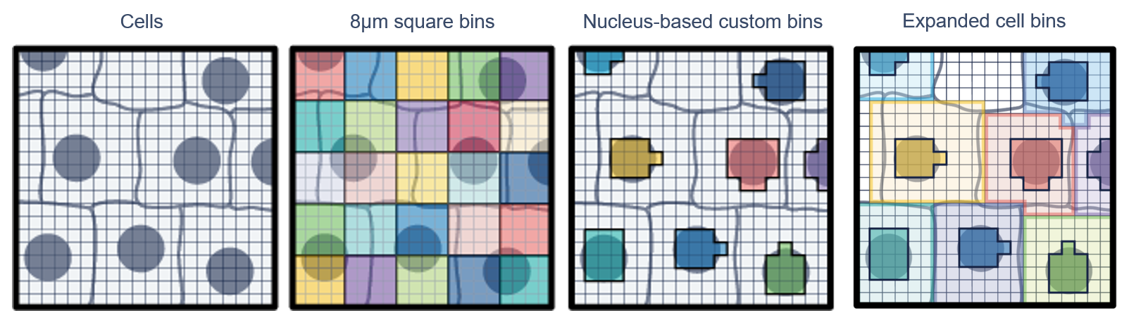
Improve cell-type resolution with nuclei expansion–based cell segmentation available in Space Ranger version 4.0 software. Importantly, Space Ranger v4.0 now supports cell and nucleus segmentation with H&E images. So you can reprocess existing Visium HD data through the new Space Ranger pipeline to derive these nuclei and cell segmentation outputs alongside the square bins
- Input a brightfield image of your H&E-stained tissue into the Space Ranger pipeline
- The software performs nuclei detection and expansion
- The Space Ranger pipeline outputs custom binning files viewable in Loupe Browser software
The new version can be found at: https://www.10xgenomics.com/support/software/space-ranger/latest
Some other notable features are:
- A new UMI-based registration algorithm for Visium HD and Visium HD 3′ data.
- You can now visualize and analyse Visium HD / Visium HD 3′ cell segmentation in Loupe Browser v9.
Watch introductory webinar on-demand
Learn more about the Visium HD 3’ assay in this introductory webinar, including a Q&A with product experts.
Build your own Visium HD 3’ experiment
Use 10x Genomics’ interactive tool to design your Visium HD 3’ experiment. Answer questions about your sample types and experimental goals, and get kit recommendations.

Other Resources
Press Release: 10x Genomics Expands Visium Platform with Launch of Visium HD 3’ Gene Expression, Unlocking Deeper Spatial Recovery
Grant application resources for Visium products
AACR Annual Meeting (Chicago) Visium HD 3′ poster: Visium HD 3’ enables unbiased whole transcriptome spatial profiling of Tumor microenvironment in fresh frozen cancer tissues at single cell scale resolution.
