About LICORbio
LICORbio offers imaging systems, reagents, software, and support for Western blots, protein gels, EMSAs, ELISAs, tissue sections, and more.
Quantitative Western Blot
A near-infrared, quantitative Western blot makes relative comparisons between different treatments possible. The goal of a quantitative Western is to accurately measure changes in protein expression.
Compatible imaging systems:
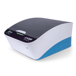
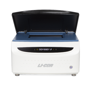
Odyssey M, Odyssey DLx

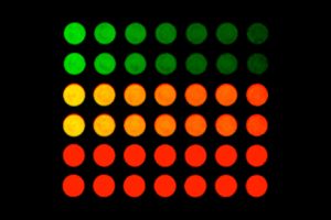
In‑Cell Western™ Assay
A quantitative immunofluorescence assay performed in multi-well plates. Data analysis is based on whole-well fluorescence quantification. This assay offers a convenient alternative to Western blotting and is a powerful platform for meaningful in situ analyses.
Compatible imaging systems:
Odyssey M, Odyssey DLx
Chemiluminescent Western Blots
Enhanced chemiluminescence (ECL) or chemiluminescent Western detection is a traditional method for conducting Western blotting research. A distinguishing characteristic is the use of an enzymatic reaction for protein detection.
Compatible imaging systems:
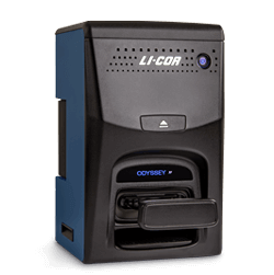
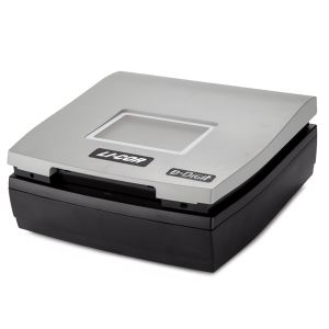
Odyssey M (Model 3350), Odyssey XF, C‑DiGit

Nuclei Acid Gels
Nucleic acid electrophoresis is a common laboratory technique that separates nucleic acids based on molecular size and structure through an agarose gel with the aid of an electric current.
Compatible imaging systems:
Odyssey M, Odyssey DLx, Odyssey XF, D‑DiGit

Protein Estimation Assays
Before beginning a Western blot experiment, one of the first things you will need to know is how much sample to load in each lane. The only way to be sure that you are loading a consistent amount of protein is to perform a protein estimation assay first. Consistent loading is critical for meaningful normalisation.
Compatible imaging systems:
Odyssey M
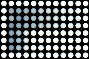
Luminescence-Based Multi-Well Plate Assays

EMSA/Gel Shift Assay
The EMSA (electrophoretic mobility shift assay) is used to study protein:DNA complexes and interactions. Protein:DNA complexes migrate more slowly than unbound linear DNA on a non-denaturing gel, causing a “shift.” Also called “gel shift” or “gel retardation” assays, the EMSA can be used to analyse sequence-specific recognition of nucleic acids by proteins.
Compatible imaging systems:
Odyssey M, Odyssey DLx

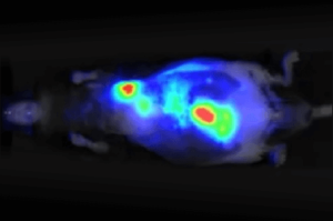
Therapeutic Delivery and Response
Optical imaging is ideal for observing and characterising delivery of your therapeutic in live animals and excised organs. Optical modalities include direct detection using near-infrared (NIR) fluorescent dyes and indirect detection using bioluminescence or NIR fluorescent proteins. NIR fluorescence detection (680 nm to 820 nm) is a sensitive and efficient modality for in vivo and ex vivo studies. It can be used to illuminate features, such as tumours and vasculature, deep within the body.

Tissue Section Imaging
Tissue section imaging is vital to advancing scientific understanding across many disciplines. Tissue section imaging is useful to study diseased tissue and to identify localisation and expression of biological targets ex vivo. Tissue sections can be used to assess therapeutic agent colocalisation, to understand diseases, and to characterise treatments. Targets can be proteins, DNA/RNA, or labelled small molecules.