
Author: karen
Millennium Science – 10x Genomics Grant Value Multiplier Program



Make your research funding go further
Planning an upcoming grant application?
The Millennium Science – 10x Genomics Grant Value Multiplier Program is designed to help Australian and and New Zealand Early and Mid Career Researchers (EMCRs) incorporate single cell and spatial biology into upcoming grant applications and get more value from their research funding.
Register your eligible grant project before submission and include 10x Genomics consumables in your proposed grant budget. If your grant is successfully awarded, you could receive an additional 50% in eligible 10x Genomics consumable value, helping you expand the scope and impact of your research.
Register your grant project using the form below:
Turn your grant funding into more research
Turn your grant funding into more research
Receive an additional 50% in eligible 10x Genomics consumable value, with no maximum cap.
The Grant Value Multiplier scales with your qualifying 10x Genomics purchase. The more eligible 10x Genomics consumables included in your successful grant, the greater the additional value you can unlock.
For example only:
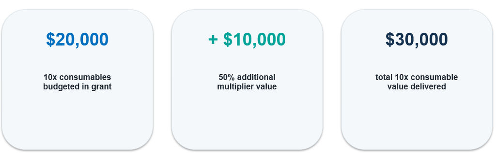
Planning $20,000 of eligible 10x Genomics consumables in your grant?
$20,000 grant budget + $10,000 Grant Value Multiplier = $30,000 total potential 10x Genomics consumable value
That additional value could help you analyse more samples, explore more biology or extend the scope of your study, without increasing the 10x Genomics budget requested in your grant.
Who is eligible?
The program is open to Australian and New Zealand researchers and research teams preparing eligible competitive research grant applications.
Early and Mid Career Researchers (EMCRs), as defined by the relevant funding scheme or institution, are particularly encouraged to apply.
You may be eligible if you:
• Are an Australian and New Zealand based researcher or research team
• Are preparing an eligible competitive research grant application
• Plan to include eligible 10x Genomics consumables in your proposed project budget
• Register your project with Millennium Science before submitting your grant application
• Proceed with the eligible 10x Genomics purchase if your grant is successfully awarded
Relevant funding opportunities may include ARC, NHMRC and MRFF schemes, as well as eligible university seed and pilot grants, philanthropic grants and disease specific research funding.
Not sure whether your grant is eligible? Register your interest and our team can help.
How it works
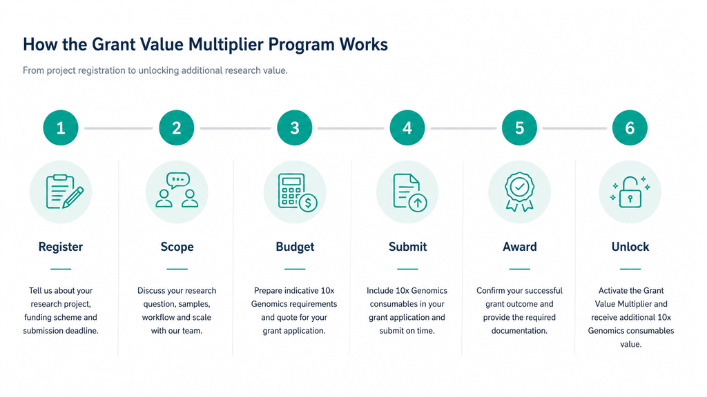
1. Register your grant project
Tell us about your research, funding scheme and submission deadline.
2. Plan your 10x Genomics component
Our team can help you scope the technology, experimental requirements and indicative budget.
3. Prepare your grant budget
Work with our team to determine indicative 10x Genomics requirements and obtain a quote for your grant application.
4. Include 10x Genomics in your grant application
Submit your application with eligible 10x Genomics consumables included in your proposed project budget.
5. Let us know when your grant is awarded
Once your successful grant and eligible purchase are confirmed, your project can proceed through the Grant Value Multiplier process.
6. Unlock additional research value
Receive additional eligible 10x Genomics consumables equivalent to 50% of the qualifying purchase value, subject to program terms and conditions.
More than a funding multiplier
You don’t need to have every detail of your experiment worked out before you register.
Before submission, Millennium Science and 10x Genomics can connect you with support to help plan the 10x Genomics component of your grant, including:
• Experimental scoping based on your research question
• Single cell and spatial technology selection
• Study design and sample planning
• Indicative consumable requirements and quotation support
• Technical resources and relevant publications
• Access to local service providers and core facilities where required
• 1:1 Grant Planning Sessions with our sales and technical teams
Our goal is to help you build a scientifically appropriate and realistically budgeted 10x Genomics component into your proposal.
Planning an upcoming grant?
Whether you’re planning your first single cell experiment, adding spatial biology to an established research program, or looking to increase the scale of your next study, start the conversation before you submit your application.
Register your grant project using the form below:
Have a project in mind but aren’t sure what to include in your grant? Request a 1:1 Grant Planning Session to discuss your project with the Millennium Science and 10x Genomics team.
Eligibility criteria, eligible products, qualifying grant schemes, purchase requirements and redemption periods apply. Participation and multiplier value are subject to approval under the final program terms and conditions. Atera reagents are excluded from the Grant Value Multiplier Program.
Agilent Liquid Handling Instruments: A Complete Guide to Readers, Washers & Automation
Agilent Liquid Handling & Microplate Instrument Portfolio: Choosing the Right Workflow Solution for Your Lab
Modern life science laboratories are under constant pressure to generate reliable data faster, improve reproducibility, and streamline increasingly complex workflows. From ELISA automation and nucleic acid quantification to high-throughput screening and live-cell analysis, selecting the right liquid handling and detection platform can significantly improve laboratory efficiency.
Agilent’s liquid handling and microplate instrumentation portfolio combines flexible automation, multimode detection, plate washing, dispensing, and workflow integration technologies designed to support research laboratories ranging from routine assay environments through to advanced high-throughput screening facilities.
Why Agilent Liquid Handling Systems?
Agilent offers a broad range of workflow solutions that help laboratories:
- Reduce manual pipetting and repetitive handling
- Improve assay consistency and reproducibility
- Scale from low-throughput to automated high-throughput workflows
- Support applications including ELISA, cell-based assays, nucleic acid quantification, microbial kinetics, phenotypic screening, and organoid workflows
- Integrate automation across washing, dispensing, detection, and plate handling
How to Choose the Right Agilent Liquid Handling Solution
The ideal platform depends on your workflow complexity, throughput requirements, and assay types.
| Workflow Need | Recommended Solutions |
| Routine ELISA workflows | 800 TS + 50 TS |
| Nucleic acid quantification | Epoch or Epoch 2 |
| Flexible multimode detection | Synergy LX or Synergy H1 |
| High-throughput screening | Synergy Neo2 + BioStack |
| Automated wash/dispense workflows | EL406 or 406 FX |
| 3D cell culture dispensing | MultiFlo FX |
| Live-cell imaging and phenotypic analysis | Cytation 9 |
Agilent Absorbance Microplate Readers
Agilent absorbance readers support routine laboratory workflows including ELISAs, protein assays, microbial growth studies, and nucleic acid quantification.
Agilent 800 TS Microplate Reader
The 800 TS is designed for straightforward absorbance workflows and cost-conscious laboratories requiring reliable performance for routine assays.
Key features
- Reads wavelengths from 400-750 nm
- Supports 6- to 384-well plates
- Touchscreen interface with USB export
- Optional temperature control and shaking
- Gen5 software integration
Best-fit applications: ELISAs, Protein assays, Enzyme kinetics, Basic cell-based assays
Agilent Epoch Microplate Spectrophotometer
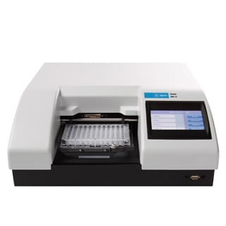
The Epoch combines monochromator-based UV-Vis detection with flexible wavelength selection for nucleic acid and protein quantification workflows.
Key features
- UV-Vis range from 200-999 nm
- Compatible with Take3 micro-volume plates
- Temperature control to 65 °C
- Supports 6- to 384-well plates
Best-fit applications: DNA and RNA quantification, 260/280 and 260/230 purity measurements, Cytotoxicity assays, Cell proliferation studies, Enzyme kinetics
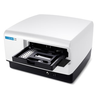
Agilent Epoch 2 Microplate Spectrophotometer
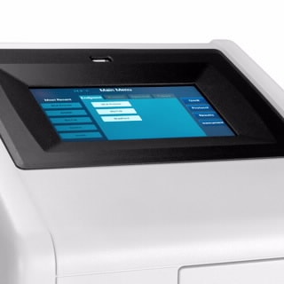
The Epoch 2 expands absorbance capabilities with stand-alone touchscreen operation and full-spectrum scanning functionality.
Ideal for: Spectral scanning, Microbial growth kinetics, Nucleic acid purity analysis, Stand-alone absorbance workflows
Automated Plate Washers for ELISA & Cell-Based Assay
Reliable washing is critical for reducing background noise and improving assay consistency in ELISA and cell-based workflows.
Agilent 50 TS Washer
The 50 TS provides dependable automated washing for routine assays and pairs naturally with absorbance readers like the 800 TS.
Applications: ELISA washing, Cell-based assays, Vacuum filtration workflows
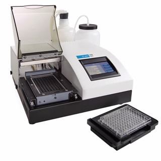
Agilent 405 LS and 405 TS Washers
These automated plate washers support more advanced workflows with dual-action manifold technology and automation compatibility.
Key features
- Automated washing for 96- and 384-well plates
- Biomagnetic separation support
- Vacuum filtration modules
- Four-buffer switching
- BioStack automation compatibility
Best-fit workflows: ELISA, Microsphere-based assays, Automated cell-based assays
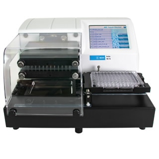
Agilent Washer-Dispenser Systems
Combining washing and dispensing into a single platform can simplify workflows and reduce instrument footprint.
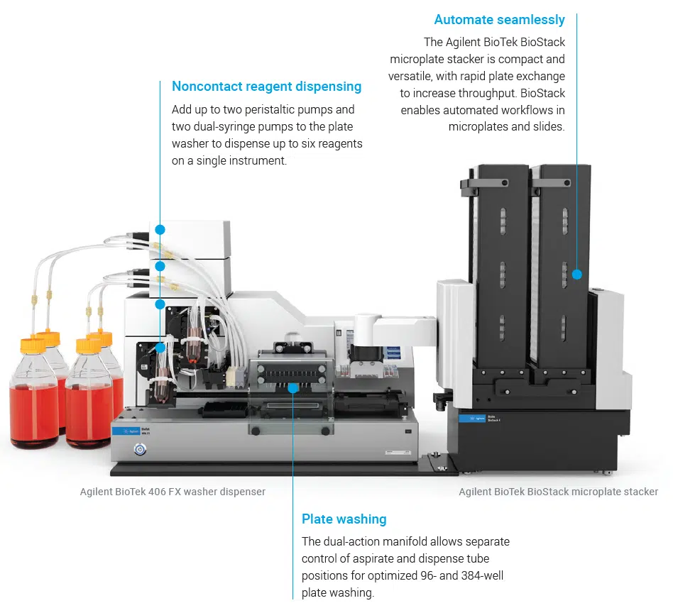
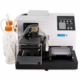
Agilent 406 FX Washer Dispenser
The 406 FX integrates washing and dispensing capabilities for automated multi-step assay workflows.
Highlights
- Washing plus up to six reagent dispensers
- Supports biomagnetic protocols
- Compatible with 96- to 1536-well plates
- Robotic integration support
Common applications: ELISA automation, Cell culture workflows, Bead-based assays
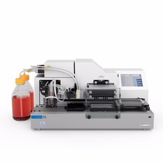
Agilent EL406 Washer Dispenser
The EL406 combines rapid washing and dispensing into a compact automation-ready platform.
Ideal for: High-throughput ELISA workflows, Multiplex assays, Integrated wash and dispense automation
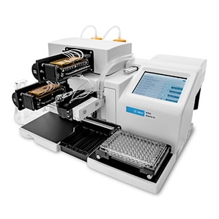
Bulk Reagent Dispensing
Agilent MultiFlo FX Multi-Mode Dispenser
The MultiFlo FX is designed for flexible reagent dispensing across a wide range of assay formats.
Key capabilities
- Dispensing to 6- to 1536-well plates
- Up to four reagents in parallel
- Gentle media exchange options for delicate cultures
Applications: 2D and 3D cell culture, Spheroid and organoid workflows, ELISA preparation, Bead-based assays
Multimode Readers for Advanced Detection
Agilent multimode readers combine absorbance, fluorescence, and luminescence detection technologies to support a broad range of assay requirements.
Agilent Synergy Neo2
The Synergy Neo2 is designed for demanding high-throughput and multiplex workflows.
Features
- Hybrid detection with filter and monochromator optics
- Laser TRF capability
- Up to four PMTs
- Environmental control with CO₂/O₂ regulation
Applications: High-throughput screening, Multiplex assays, Advanced multimode workflows
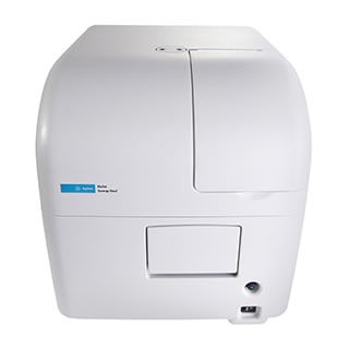
Agilent Synergy HTX
The Synergy HTX provides economical multimode detection for routine assay environments.
Detection modes: Absorbance, Fluorescence, Luminescence, Alpha assays
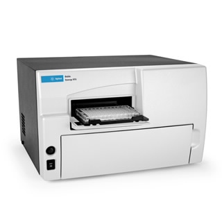
Agilent Synergy LX
The Synergy LX is designed for straightforward multimode detection with an intuitive touchscreen interface.
Ideal applications: Nucleic acid quantification, ELISA, BCA and Bradford assays, Cell viability workflows
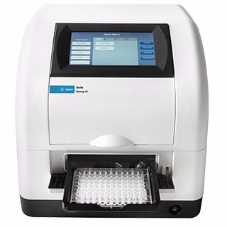
Agilent Synergy H1
The modular Synergy H1 platform enables laboratories to scale capabilities as assay requirements evolve.
Supports: Absorbance, Fluorescence, Luminescence, AlphaScreen, TRF workflows
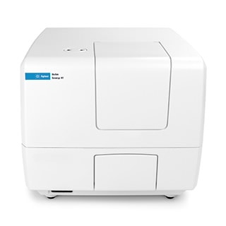
Imaging & High-Content Analysis Workflows
Agilent Cytation 9
The Cytation 9 combines automated imaging with multimode detection in a single platform.
Key capabilities
- Imaging up to 60× magnification
- Fluorescence, luminescence, and UV-Vis detection
- Live-cell environmental control to 65 °C
- BioStack automation compatibility
Common applications: Live-cell imaging, Phenotypic screening, Quantitative cell analysis
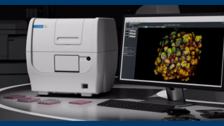
High-Throughput Laboratory Automation
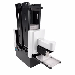
Agilent BioStack
The BioStack automates plate loading and unloading for walk-away workflow automation.
Benefits
- Fast plate exchange
- Delidding and re-lidding support
- Compatible with 6- to 1536-well plates
- Enables unattended high-throughput processing
Supporting Modern Research Workflows
Agilent’s liquid handling and microplate instrumentation portfolio supports a wide spectrum of laboratory applications, from routine absorbance assays through to advanced automation and high-content workflows. With scalable solutions for washing, dispensing, detection, imaging, and plate automation, laboratories can build flexible workflows that improve reproducibility, efficiency, and throughput.
To discuss the best Agilent liquid handling workflow for your laboratory, contact the team at Millennium Science.
On this page
- Agilent Liquid Handling & Microplate Instrument Portfolio: Choosing the Right Workflow Solution for Your Lab
- Why Agilent Liquid Handling Systems?
- How to Choose the Right Agilent Liquid Handling Solution
- Agilent Absorbance Microplate Readers
- Automated Plate Washers for ELISA & Cell-Based Assay
- Agilent Washer-Dispenser Systems
- Bulk Reagent Dispensing
- Multimode Readers for Advanced Detection
- Imaging & High-Content Analysis Workflows
- High-Throughput Laboratory Automation
- Supporting Modern Research Workflows
10x Genomics Clinician Researcher Accelerator Program
10x Genomics Clinician Researcher Accelerator Program
Clinical research is increasingly driven by the need to understand biological heterogeneity, identify actionable biomarkers, and link molecular mechanisms to patient outcomes. Traditional bulk and low-resolution approaches often mask clinically relevant signals, slowing discovery and translation. 10x Genomics technologies accelerate clinical research by enabling cell-resolved and spatially resolved insights at scale at earlier in the research pipeline.
A targeted Clinician Researcher Accelerator Program designed to support medically qualified clinician researchers in generating high-impact spatial and single-cell data, with dedicated support and preferential pricing to fast-track translational research.
The Clinician Researcher Accelerator Program is a limited-time initiative supporting medically qualified clinician researchers who are looking to integrate spatial transcriptomics and single-cell profiling into their research. Successful applicants will receive access to dedicated technical guidance, and preferential pricing to help generate robust pilot data for translational and clinically focused studies.
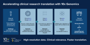
1. Resolving clinical heterogeneity at single-cell resolution
Many clinical samples including tumours, immune tissues, and biopsies, are highly heterogeneous. Bulk profiling averages signals across cell populations, obscuring rare or disease-driving cell states.
Chromium Single Cell technologies enable researchers to:
- Identify rare or pathogenic cell populations
- Characterise immune cell diversity and activation states
- Study patient-to-patient variability at cellular resolution
Clinical impact:
Earlier identification of clinically relevant subpopulations improves hypothesis generation, patient stratification strategies, and biomarker discovery.
2. Linking molecular signals to tissue context
Understanding where cells are located within tissue is critical for clinical interpretation, particularly in oncology, immunology, and pathology-driven research.
Visium Spatial Gene Expression allows researchers to:
- Map gene expression within intact tissue sections
- Correlate molecular patterns with histology
- Identify spatially distinct disease niches
Clinical impact:
Spatial context strengthens biological interpretation, supports translational relevance, and enables more compelling mechanistic narratives for grants and publications.
3. Translating discovery into spatial validation
Discovery technologies are most powerful when paired with targeted validation in clinically relevant samples.
Xenium In Situ enables:
- High-plex, single-cell resolution spatial validation
- Direct visualisation of clinically relevant gene signatures
- Confirmation of biomarkers within preserved tissue architecture
Clinical impact:
Xenium supports the transition from discovery to validation which is a critical step for translational studies, diagnostic development, and clinical adoption.
4. Accelerating the path from pilot data to funding
Clinical research is often constrained by:
- Limited sample availability
- Ethical and governance considerations
- The need to justify scale and cost upfront
10x Genomics platforms are well suited to small, high-information pilot studies, allowing researchers to:
- De-risk experimental design
- Generate convincing preliminary data
- Strengthen ARC, NHMRC, and translational grant applications
Clinical impact:
High-quality pilot data reduces uncertainty and accelerates progression to larger, funded studies.
5. Enabling multi-modal clinical insights
Complex clinical questions rarely have single-dimensional answers.
10x Genomics workflows support:
- Integration of transcriptomics, immune profiling, and spatial biology
- Cross-platform studies linking cell state, location, and function
- More comprehensive biological models of disease
Clinical impact:
Multi-modal data improves robustness, reproducibility, and translational relevance which is increasingly expected by reviewers and collaborators.
6. Supporting reproducibility and scalability in clinical settings
Clinical research demands consistency, robustness, and scalability.
10x Genomics technologies are:
- Highly standardised and widely adopted
- Supported by validated workflows and protocols
- Designed for reproducible data generation across sites
Clinical impact:
Standardisation supports multi-centre studies and facilitates collaboration between hospitals, academia, and industry.
Apply for the Clinician Researcher Accelerator Program now!
- One winner will be awarded 50% discount across Xenium v1 or 5k Prime panel*(2 slides) and Xenium running consumables Or One Flex kit (16-sample configuration)
- All approved applicants will receive a 25% discount
- Orders must be placed by 19 June 2026
*Custom panels excluded.
Application timeline
• Applications close: 27 May 2026
• Outcome notification: 1 June 2026
Fill in the form below to submit your application now!
On this page
- 10x Genomics Clinician Researcher Accelerator Program
- 1. Resolving clinical heterogeneity at single-cell resolution
- 2. Linking molecular signals to tissue context
- 3. Translating discovery into spatial validation
- 4. Accelerating the path from pilot data to funding
- 5. Enabling multi-modal clinical insights
- 6. Supporting reproducibility and scalability in clinical settings
- Apply for the Clinician Researcher Accelerator Program now!
2026 Millennium Science – 10x Genomics Fellowship Program
Special focus edition: FFPE
Who This Fellowship Is For
Graduate students and early to mid-career researchers with no prior hands-on experience using 10x Genomics technologies are invited to apply for the prestigious 2026 Millennium Science – 10x Genomics Fellowship Program.
This Fellowship is designed to support researchers who are keen to adopt cutting-edge single-cell and spatial technologies in their work.
Important Dates
Applications close: 16 March 2026
Outcomes announced: 30 March 2026
Fellowship Focus: FFPE Samples
The 2026 Fellowship Program focuses on the application of 10x Genomics technologies to FFPE samples, using one or more of the following platforms:
Chromium Single Cell
Visium Spatial
Xenium In Situ (NEW within the Fellowship Program!)
Fellowship Benefits
Successful applicants will receive:
- A personalised mentoring session on experimental design with a 10x Genomics specialist
- Dedicated support from the Fellowship Program Coordinator
- 10x Chromium or Visium CytAssist demo instrument placement (if required and subject to availability)
- Support with project onboarding through a core facility running 10x Genomics platforms (Chromium, Visium or Xenium)
- Discounted 10x Genomics consumables*
- Invitation to present at Millennium Science–10x Genomics events
- Membership in the Millennium Science–10x Genomics Fellows Network
- Fellowship-exclusive merchandise
- A certificate confirming completion of the Fellowship Program
Applications will be assessed by a panel of scientific experts from Millennium Science and 10x Genomics, based on project innovation, feasibility, and potential to inspire future research.
Through this program, Millennium Science and 10x Genomics aim to empower early and mid-career researchers to generate high-quality pilot data using advanced multi-omics technologies, supporting future experimental design and funding applications.
*Please note, the Fellowship is not associated with provision of free reagents. Discounts cannot be combined with other promotions.
Application Details
To apply, please submit:
- Project title: 20 words maximum
- Abstract: 250 words maximum
- CV: Attach 1-page CV
- Letter of Support: Please provide a letter of support from the principal investigator of the laboratory. Additional institutional or departmental letters of reference may be submitted to support the application.
Completion of the online application survey below is required to submit your application.
Applications will be reviewed by Millennium Science as they are being received. Successful applicants will be notified via email.
Conditions of Eligibility
- No previous personal experience using 10x Genomics technologies
- Open to graduate students and early to mid-career researchers (up to 15 years post-PhD; special circumstances may be considered with supporting documentation)
- Selected candidates must complete the specified individualised training program prior to reagent delivery or instrument placement
- Applicants must be based in Australia or New Zealand
Hear from Our 10x Genomics Fellowship Alumni

“I was fortunate to receive the Millennium Science-10x Genomics Fellowship in 2021 during my first postdoctoral appointment in Associate Professor Megan Wilson’s lab at the Department of Anatomy, University of Otago, where her lab studies whole-body regeneration in the marine tunicate Botrylloides diegensis. At a time when single-cell technologies were still emerging, the Fellowship enabled us to rapidly adopt 10x Genomic technology and interrogate this process at unprecedented resolution. After some optimisation, our first 10x run produced a high-quality dataset that underpinned a recent publication in Development. Importantly, the Fellowship established long-term single-cell capability in the lab, directly benefiting lab member Berivan Temiz, who applied this method extensively during her PhD. I continue to find 10x Genomics the most user-friendly and straightforward to implement.” Michael Meier
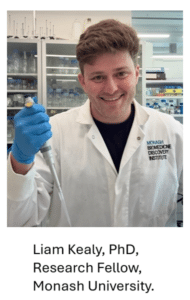
“I moved to Melbourne in 2015 from Phuket, Thailand to pursue my passion for biomedical science. I completed my PhD at Monash University in 2023,investigating how epigenetic mechanisms shape B-cell biology, particularly the role of the histone methyltransferase DOT1L in regulating immune cell identity and function. My research is driven by a desire to understand how immune cells adapt in different disease contexts and how gene regulation disruptions contribute to chronic inflammatory conditions. In early 2023, I was honoured to receive the Millennium Science-10x Genomics Fellowship, which has been pivotal in advancing this work. With their support, I implemented single-cell multiome sequencing to profile circulating B cells from individuals with chronic disease, including Long COVID-a debilitating, multi-system syndrome that remains poorly understood. By measuring gene expression and chromatin accessibility in the same cell, this approach reveals subtle immune signatures and hidden cellular states, opening new avenues for diagnosis and treatment. The Fellowship provided not only advanced technology, but also personalised mentorship, technical guidance, and connection to a collaborative network of fellows across Australia and New Zealand. This support has accelerated my research and positioned me to tackle ambitious questions about immune dysfunction in complex diseases like Long COVID.” Liam Kealy
2026 Information Session
Any questions?
If you have any questions, please contact us at fellowship@mscience.com.au.
Beyond Flow Cytometry: Unlocking Single-Cell Insights with 10x Genomics
Bridging Flow Cytometry with Single-Cell Sequencing
Flow cytometry has long been the backbone of high-dimensional single-cell analysis. With the ability to measure up to ~45 parameters per cell, flow has enabled researchers and core facilities to rapidly phenotype complex populations and enrich rare cell types with confidence.
But what if you could go further — without disrupting your existing workflows?
By integrating flow cytometry with single-cell sequencing, researchers can now move beyond predefined panels and surface markers to uncover deeper, unbiased biological insight.
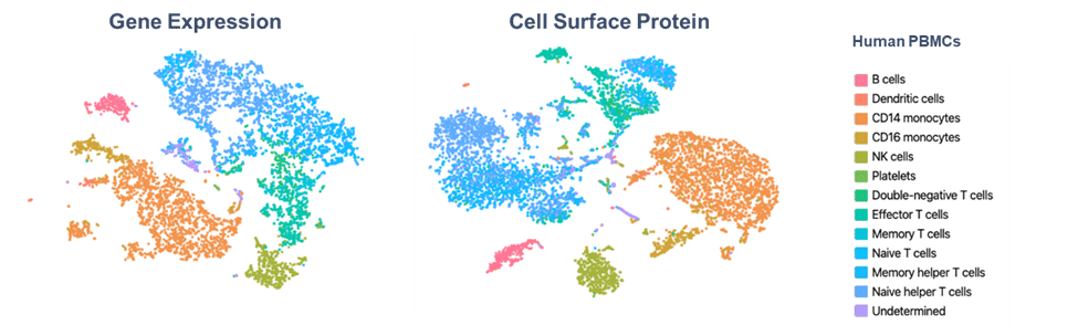
Why Bridge Flow Cytometry with Single-Cell Sequencing?
Flow cytometry has long been the gold standard for measuring what you expect to see. But what about the biology you don’t expect? What about the rare cell states, subtle activation programs, and novel biomarkers that surface markers alone simply can’t capture?
This is where single-cell sequencing transforms your capabilities. By bridging flow cytometry with the 10x Genomics platform, you can move from measuring dozens of parameters to profiling 300+ protein markers—with the option to simultaneously capture whole-transcriptome gene expression from the same single cell.
Unmatched multiplexing without compromise
Breaking free from spectral overlap limitations opens new possibilities. Instead of carefully balancing fluorophore combinations, you can now interrogate hundreds of protein markers alongside comprehensive gene expression data. This quantum leap in multiplexing reveals cellular complexity that traditional flow simply cannot access.
Discovering What’s Hidden
Many critical cell states appear identical by surface protein analysis alone. Activation, exhaustion, differentiation, and stress states often require deeper molecular investigation to truly distinguish. Single-cell sequencing uncovers these hidden layers of heterogeneity, revealing novel populations and functional states that would otherwise remain invisible.
Deeper biology from enriched populations
Leverage FACS to isolate rare or complex populations, then apply single-cell sequencing to achieve transcriptome-wide and high-plex proteomic resolution.In short: sort with flow, then profile with depth.
Designed for Your Workflow
The beauty of this approach is that it builds on what you already do exceptionally well. Use FACS to enrich the rare or complex populations you’re interested in, then apply single-cell sequencing to achieve transcriptome-wide resolution. Your expertise in flow cytometry becomes the foundation for even more powerful discoveries.
What Can You Unlock?
By pairing flow cytometry with single-cell sequencing, researchers can access a new layer of biological resolution:
-
Unbiased cell type and state discovery
Identify known and novel populations without relying on predefined antibody panels. -
Protein validation with gene expression
Confirm antibody signals and distinguish protein presence from true pathway activation. -
Functional state resolution beyond surface markers
Dissect activation, exhaustion, differentiation and stress states that appear identical by flow. -
Pathway-level biology at scale
Interrogate canonical pathways such as Wnt, TCR and interferon signalling through coordinated gene expression, rather than single proxy markers.
New Flexible and Cost-Effective Options
With the introduction of Single Cell Flex v2.0 (Protein-Only), high-plex single-cell protein profiling is now more accessible than ever.
This flexible option enables:
-
High-plex protein analysis at a significantly reduced cost
-
Support for more projects, more users and more samples
-
No requirement for RNA profiling, where transcriptomics isn’t needed
For many flow cores, this opens the door to offering advanced single-cell services without the overhead of full transcriptome assays.
The Natural Next Step for Flow Cytometry
The integration of 10x Genomics technology with your existing flow cytometry services doesn’t mean disrupting established workflows—it means enhancing them. Your team’s expertise in high-dimensional single-cell analysis positions you perfectly to take this next step.
As the authorised 10x Genomics distributor in Australia and New Zealand, Millennium Science is here to help you understand how single-cell sequencing can complement and extend your current offerings. Whether you’re looking to add new capabilities, support cutting-edge research, or simply explore what’s possible beyond traditional flow, our specialists are ready to guide you through the options.
Ready to expand your core’s capabilities?
Contact our team today to learn more about 10x Genomics solutions and how they can seamlessly integrate into your core facility workflows.
2025 End of Year Deliveries & Holiday Schedule
Important Ordering Dates
To help us meet your delivery requirements before the Christmas break, please note the following important dates:
- For general consumable items: Place your order by 12 PM, Wednesday 26 November 2025.
- For 10x Genomics consumables: Place your order by 4 PM, Monday 1 December 2025.
- Instrument delivery times will be provided on a case-by-case basis.
Orders received after these dates are welcome, however delivery will likely take place in early 2026 due to expected closure of institute receiving facilities.
Holiday Schedule
- Our office will close at 12 PM on Tuesday 23 December 2025 and reopen on Friday 2 January 2026.
- No orders will be processed during the office closure.
- The Millennium Science warehouse’s final shipping day will be Wednesday 17 December 2025, resuming dispatches on Monday 5 January 2026.
Thank you for your continued support throughout 2025. We wish you a safe and joyful holiday season!
Please note: These dates are subject to product availability and the delivery facilities at your institution being operational.
On this page
Meet EYRA: Multiplexing Reimagined
A Joe Blogs post by Joe Roberts, PhD
When it comes to multiplex protein analysis, researchers need accuracy, speed, and simplicity. Traditional flow-based systems have long been the standard, but they bring challenges: sheath fluids, blocked probes, and constant maintenance. Enter the Mabtech EYRA™ – a fluidics-free multiplex immunoassay platform that reimagines how scientists generate cytokine and biomarker data. With confocal imaging, RAWsphere analysis, and compatibility with EYRAplex bead kits, EYRA makes multiplexing faster, simpler, and more reliable.
Multiplex Without Compromise
With EYRAplex magnetic bead assays, EYRA can quantify more than 30 analytes from a single sample – whether that’s serum, plasma, or cell culture supernatant. This means less sample consumption, fewer runs, and richer datasets for every experiment.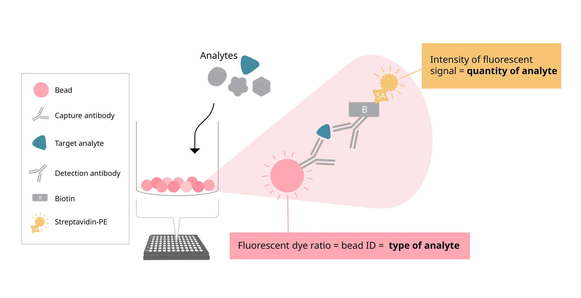
At the core of EYRA’s precision is the RAWsphere image analysis algorithm. It identifies each bead, links it to the correct analyte, and quantifies the PE signal with high resolution. Whether you’re measuring six cytokines or a full 30+ panel, RAWsphere ensures your multiplex data is accurate and reproducible.
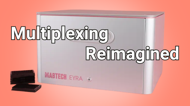
Watch video to see how EYRA works
Fluidics-Free Technology
EYRA is built around a completely flow-free design. No sheath fluid, priming, waste handling or blocked probes.
Instead of pushing samples through fluidics, EYRA uses confocal microscopy to image settled magnetic beads directly in the wells of a 96-well plate. Each bead carries a unique fluorescent dye signature for identification, while analyte-bound PE-labelled antibodies provide the quantitative signal.
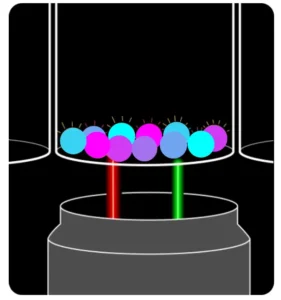
The result? You insert your plate, select your assay in the intuitive Mabtech Opal™ software, and hit read. Fifteen minutes later, you have fully processed data – with your samples never leaving the wells.
From Plate to Excel - Fast
Opal™ software comes preloaded with templates for every EYRAplex kit. Standards, plate layouts, and gating are handled automatically – so you spend less time setting up and more time on results.
Once the plate read is complete, results are exported directly into Excel, with options for bulk export if you’re working on large studies or multiple plates. No manual reformatting. No data wrangling headaches.
The Maintenance-Free Mindset
Because EYRA is fluidics-free, there’s no daily calibration or cleaning. No flushing, no wasted consumables, and no time lost to instrument downtime. It’s genuinely a plug-and-play experience – switch it on, run your plate, and walk away with your data.
Joe’s Takeaway
The Mabtech EYRA isn’t just another multiplex platform – it’s multiplexing reimagined. By removing fluidics and harnessing high-resolution confocal imaging, EYRA delivers:
- High-plex capacity: 30+ analytes per well
- Fast turnaround: ~15 minutes per plate
- No maintenance: no daily cleaning or calibration
- Accurate, reproducible results: powered by RAWsphere analysis
For labs looking to streamline their workflow without sacrificing data quality, EYRA offers a fresh, frustration-free alternative to traditional flow-based systems.
Until next time… happy experimenting!
Joe Roberts, PhD
Product Manager
Millennium Science

Spot the Difference: ELISA vs ELISpot vs FluoroSpot
A Joe Blogs post by Joe Roberts, PhD
When it comes to measuring proteins such as cytokines, antibodies, or growth factors, there’s no shortage of options. ELISA, ELISpot, and FluoroSpot all have important roles to play. While each assay relies on antibody-based detection, knowing which one to use (and when) can mean the difference between simply collecting data and gaining real insight.
As a Product Manager at Millennium Science, I speak with researchers every day who are deciding between these techniques. In this blog we will explore each assay to help you spot the difference and help you choose the right assay for your next experiment.
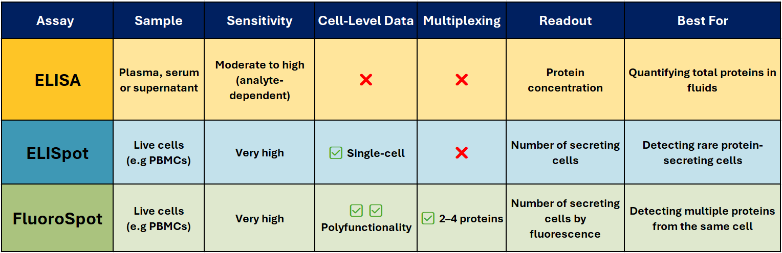
Comparison at a Glance – ELISA vs ELISpot vs FluoroSpot
ELISA – The Trusted Workhorse 📊
Best for: Quantifying total soluble protein in biological fluids.
ELISA (Enzyme-Linked Immunosorbent Assay) is one of the most widely used immunoassays for detecting and quantifying soluble proteins, such as cytokines, antibodies, and hormones, in serum, plasma, or cell culture supernatants. It’s robust, scalable, and ideal for high-throughput analysis when you need accurate, reproducible concentration data.
How it works (Sandwich ELISA):
A capture antibody is coated onto a high-binding plate and binds the target protein in your sample. A biotinylated detection antibody binds a different epitope, followed by a streptavidin–enzyme conjugate. Addition of a colourimetric substrate produces a measurable signal proportional to protein concentration.
Why researchers choose it:
- Delivers quantitative results (e.g., pg/ml or ng/ml)
- Compatible with high-throughput and automation
- Well-established, with widely available equipment
- Ideal for comparative analysis across multiple samples
Limitations:
- No information on the number or type of cells producing the protein
- Sensitivity can be lower than single-cell assays
ELISpot – Spotlight on Secreting cells
Best for: Counting individual protein-secreting cells.
ELISpot (Enzyme-Linked ImmunoSpot) detects and counts cells that secrete a specific protein. It’s particularly valuable when studying immune responses where the frequency of antigen-specific T or B cells is low – for example in vaccine research, oncology, and autoimmune disease studies.
How it works:
Live immune cells are added to a PVDF plate pre-coated with a capture antibody. When stimulated, the cells secrete the target protein, which is immediately bound near the cell. After washing away cells, a detection antibody and enzyme conjugate are added. A precipitating substrate forms a visible spot at each secretion site – each spot representing a single responding cell.
Why it stands out:
- Extremely sensitive – can detect one responder cell in >100,000
- Functional readout of immune activity
- Ideal for rare antigen-specific responses
- Widely used in T-cell response monitoring, vaccine trials, allergy research, and immuno-oncology
Pairing with Mabtech’s ASTOR2 automated ELISpot reader delivers rapid, high-precision spot counting with consistent, reproducible results across entire assay plates, and when used with Mabtech monoclonal antibody pairs and ready-to-use ELISpot kits, ensures reliable performance across species and sample types.
FluoroSpot – Multiplex Made Easy 🌈
Best for: Analysing polyfunctional immune responses.
FluoroSpot builds on the ELISpot principle but uses fluorescently labelled detection antibodies to identify multiple proteins secreted by the same cell – typically 2-4 proteins. This allows you to measure not just whether a cell responds, but how it responds.
Why it’s powerful:
- Detects polyfunctionality – cells secreting multiple cytokines
- High sensitivity with low background
- Enables simultaneous detection of different functional subsets
- Excellent for vaccine research, infectious disease, and cancer immunotherapy
Pairing with Mabtech’s IRIS2 ELISpot, FluoroSpot and FociSpot reader enables precise, high-resolution spot detection across multiple fluorescence channels.
Joe’s Takeaway
- ELISA → When you want to know how much protein is present in a sample.
- ELISpot → When you need to know how many cells are producing a protein.
- FluoroSpot → When you need to know which protein a cell is producing, and whether it’s producing more than one at the same time.
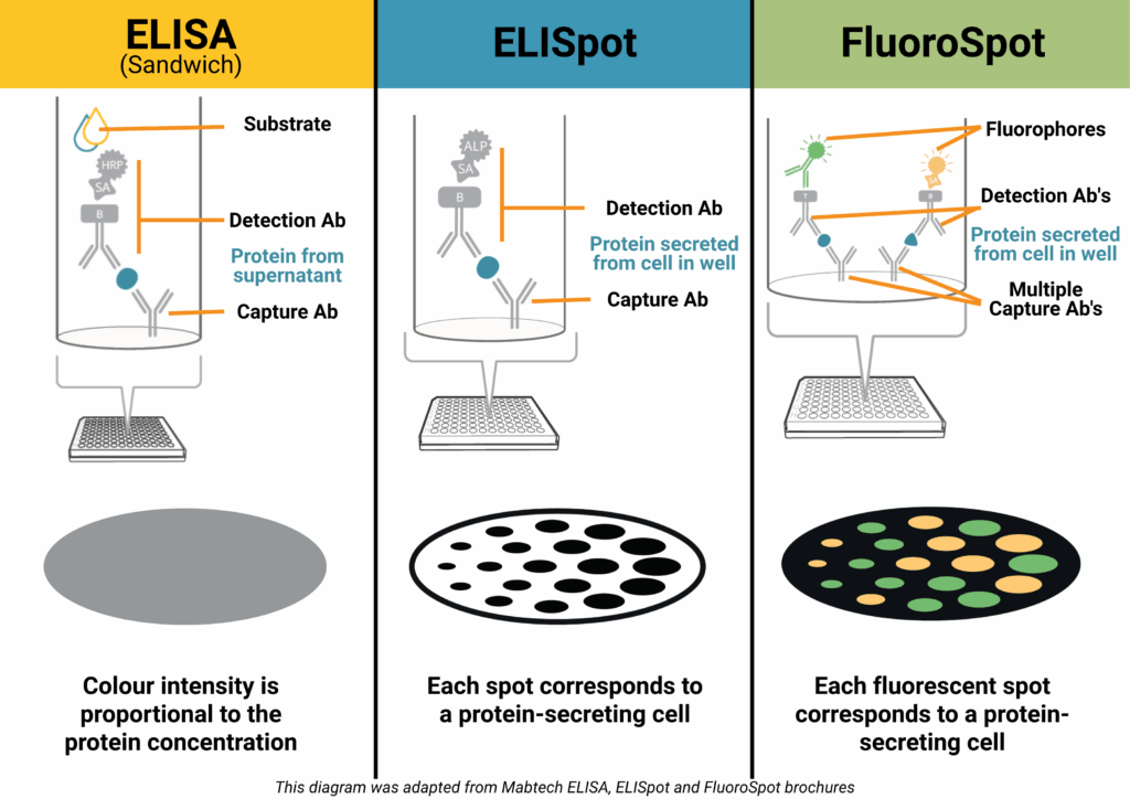
In many projects, using ELISA and ELISpot/FluoroSpot together can give the clearest picture — combining quantitative bulk measurements with functional single-cell insights.
If you’d like help selecting the right assay for your research, get in touch with our team at Millennium Science. We’re local experts, and Mabtech’s range covers everything from research use to clinical-grade applications.
Until next time… happy experimenting!
Joe Roberts, PhD
Product Manager
Millennium Science
Revolutionising RNA purification with Automation
Accelerating molecular workflows has never been easier
Norgen Biotek’s Magnetic Bead-Based Total RNA Purification Kit is now fully automated, making high-throughput RNA extraction faster, more reliable, and easier to scale.
Designed to isolate all RNA sizes – including microRNA and small RNA – with exceptional purity and reproducibility, this kit delivers consistent performance across a wide range of sample types. Whether you’re working with tissues, cells, bacteria, viruses, bodily fluids, plants or fungi, Norgen Biotek’s advanced RNA purification technology ensures high-yield results without compromising quality.
Save Time with Automated RNA Purification
Manual RNA extraction can be labour-intensive and prone to variability. Norgen Biotek’s magnetic bead-based kit streamlines the process, reducing hands-on time to just 15 minutes, while enhancing consistency across runs.
Built for Lab Automation Platforms
This kit is fully compatible with leading lab automation platforms, including:
-
Opentrons Flex – a modular and cost-effective automation system designed to scale with your lab.
-
Tecan
-
Thermo Fisher KingFisher
-
Hamilton
-
IsoPure
Seamless integration allows labs to boost throughput, minimise human error, and free up valuable time for high-value analysis and discovery.
Why Choose Norgen Biotek RNA Kits?
✅ Compatible with a broad range of sample types
✅ High-quality RNA including microRNA and small RNA
✅ Automation-ready for reliable scalability
✅ Reduced hands-on time and increased reproducibility
Interested in upgrading your RNA workflows?
Explore how Norgen Biotek and Opentrons can modernise your lab’s RNA extraction today or contact Narisa Dawar our Norgen Biotek Product Manager – ndawar@mscience.com.au.
MedChemExpress Anti-Cancer Compound Library
MedChemExpress (MCE) is a supplier of high-quality chemicals, biochemicals and compound libraries, supporting the global scientific research community. MCE’s extensive product portfolio includes a range of screening libraries designed to accelerate drug discovery and development across multiple therapeutic areas. Among these, the Anti-Cancer Compound Library is a powerful tool for researchers seeking targeted solutions and novel cancer therapies.

The library offers over 8,000 compounds with activity against both solid and haematologic tumours, making it suitable for high-throughput screening (HTS) and high-content screening (HCS).
This collection spans key cancer-related pathways such as apoptosis, cell cycle regulation and signal transduction. It also covers diverse signalling pathways — including kinase, GPCR and epigenetics — providing researchers with a comprehensive platform to explore and identify potential anti-cancer agents.
Many of the compounds have undergone rigorous clinical or preclinical evaluations, with some already approved by the FDA, highlighting their therapeutic potential.
The bioactivity and safety profiles of the compounds make the library a useful resource for advancing cancer research. Each compound is also validated for high purity and quality using NMR and LC/MS techniques, which should lead to precise and reproducible results.
A key advantage of MCE’s compound libraries is that users can fully customise a library to meet research needs from the type, quantity and concentrations of compounds and layout, offering the flexibility that researchers need to drive meaningful discoveries in oncology.
Click here to download the MedChemExpress Compounding Libraries brochure or contact Millennium Science at customerservice@mscience.com.au.